. 2012;7(3):e33539.
doi: 10.1371/journal.pone.0033539. Epub 2012 Mar 28.

The ontogenetic osteohistology of Tenontosaurus tilletti

Affiliations

The ontogenetic osteohistology of Tenontosaurus tilletti

Sarah Werning. PLoS One. 2012.

Abstract

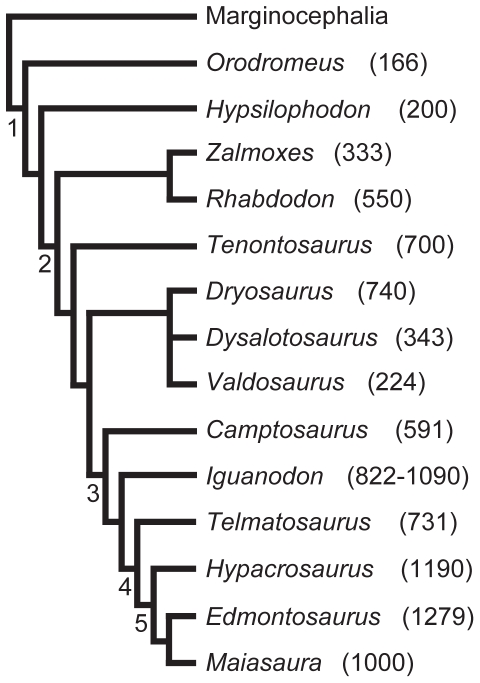
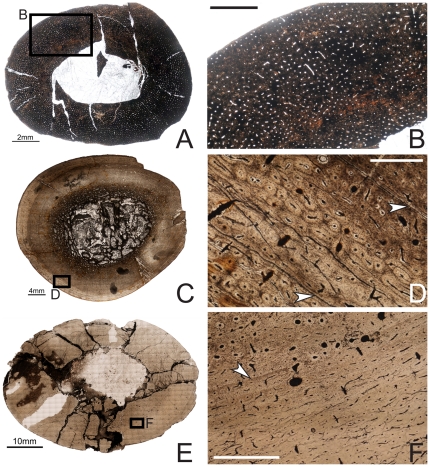
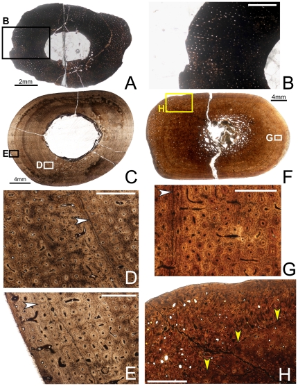
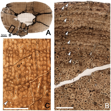
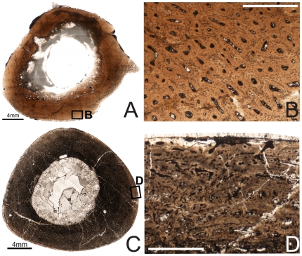
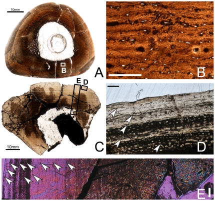
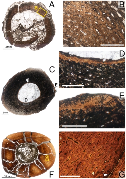
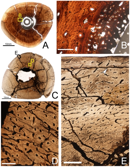
Tenontosaurus tilletti is an ornithopod dinosaur known from the Early Cretaceous (Aptian-Albian) Cloverly and Antlers formations of the Western United States. It is represented by a large number of specimens spanning a number of ontogenetic stages, and these specimens have been collected across a wide geographic range (from central Montana to southern Oklahoma). Here I describe the long bone histology of T. tilletti and discuss histological variation at the individual, ontogenetic and geographic levels. The ontogenetic pattern of bone histology in T. tilletti is similar to that of other dinosaurs, reflecting extremely rapid growth early in life, and sustained rapid growth through sub-adult ontogeny. But unlike other iguanodontians, this dinosaur shows an extended multi-year period of slow growth as skeletal maturity approached. Evidence of termination of growth (e.g., an external fundamental system) is observed in only the largest individuals, although other histological signals in only slightly smaller specimens suggest a substantial slowing of growth later in life. Histological differences in the amount of remodeling and the number of lines of arrested growth varied among elements within individuals, but bone histology was conservative across sampled individuals of the species, despite known paleoenvironmental differences between the Antlers and Cloverly formations. The bone histology of T. tilletti indicates a much slower growth trajectory than observed for other iguanodontians (e.g., hadrosaurids), suggesting that those taxa reached much larger sizes than Tenontosaurus in a shorter time.


Conflict of interest statement

Competing Interests: The author has declared that no competing interests exist.

Figures

Figure 1
Figure 1. Phylogenetic relationships and adult femoral lengths (mm) of ornithopod taxa discussed in this study.
This tree is pruned from recently published estimations of ornithischian and ornithopod , dinosaurs. These studies do not conflict in their assessment of relationships of these taxa. Node numbers correspond to the following clades: 1 – Ornithopoda, 2 – Iguanodontia, 3 – Ankylopollexia, 4 – Hadrosauroidea, 5 – Hadrosauridae. Femoral lengths are taken from , except Zalmoxes and Maiasaura .
Figure 2
Figure 2. Osteohistology of the mid-diaphyseal humerus of Tenontosaurus in a juvenile (A, B), subadult (C, D) and adult (E, F).
A. Cross-section of OMNH 10144. This bone was invaded by bacteria before fossilization and thus much of the primary tissue is obscured. It is presented here in cross-section to illustrate vascular density and arrangement. B. Detail of A, showing general vascular patterning. The cortex is dominated by longitudinal canals arranged circumferentially. C. Cross-section of OMNH 8137. D. Detail of C, showing primary cortical tissues. The bone is woven, and most canals are longitudinal primary osteons (some anastomose circumferentially). Two LAGs (arrows) are shown. E. Cross-section of FMNH PR2261. F. Detail of E, showing mostly primary tissues of the midcortex at a transition to slower growth. Deeper in the cortex (upper left), bone is woven and osteocytes are dense and disorganized. Some secondary osteons are visible, but they do not overlap or obscure all of the primary tissues. Past the LAG (arrow), canals remain dense but decrease in diameter, bone tissue is weakly woven, and osteocytes decrease in number and become more organized. Scale bars: A = 2 mm; B, F = 1 mm; C = 4 mm; D = 0.5 mm; E = 10 mm.
Figure 3
Figure 3. Osteohistology of the mid-diaphyseal ulna of Tenontosaurus in a juvenile (A, B) and two subadults (C–E, F–H).
A. Cross-section of OMNH 10144. This bone was invaded by bacteria before fossilization and thus much of the primary tissue is obscured. It is presented here in cross-section to illustrate vascular density and arrangement. B. Detail of A, showing general vascular patterning. The cortex is dominated by longitudinal canals arranged radially. C. Cross-section of OMNH 2531. D. Detail of the midcortex of C, showing primary cortical tissues. Longitudinal primary osteons are visible in primary woven bone tissue. One LAG (arrow) is shown. E. Detail of the periosteal region of C, showing the surface condition in ulnae that lack the middiaphyseal rugosity. Longitudinal primary osteons are visible in primary woven bone tissue, very similar to the tissue of the midcortex. F. Cross-section of OMNH 34191. G. Detail of the midcortex of F, showing primary cortical tissues. Midcortical tissues are similar to those shown in OMNH 2531. One LAG (arrow) is shown. H. Detail of the periosteal region of F, showing the periosteal condition in ulnae with a rugosity on the posterior surface of the bone at midshaft. The histology of this rugosity is a strongly woven, high vascularized, and very disorganized tissue that builds along a surface similar to that seen in E (yellow arrows). It grades laterally into and is capped by more typical primary bone tissue identical to that of the rest of the cortex. Scale bars: A = 2 mm; B = 1 mm; C,F = 4 mm; D,E,G = 0.5 mm; H = 2 mm.
Figure 4
Figure 4. Osteohistology of the mid-diaphyseal ulna of Tenontosaurus in two adults.
A. Cross-section of FMNH PR 2261. B. Detail of A, showing primary and secondary tissues of the mid- and outer cortex. The midcortex experiences dense secondary remodeling, and the outer cortex shows longitudinal primary osteons in parallel-fibered bone tissues grading into longitudinal simple canals in lamellar bone. Seven LAGs (arrows) are shown. C. Detail of OMNH 62990 (no cross-section shown), illustrating the radial arrangement of longitudinal primary osteons in an unremodeled area of the midcortex. One LAG (arrow) is shown. Periosteal surface to the top of this image. Scale bars: A = 4 mm; B,C = 1 mm.
Figure 5
Figure 5. Osteohistology of the diaphyseal femur of Tenontosaurus in two juveniles.
A. Cross-section of MOR 679. This section is slightly more proximal compared to others in this sample and shows part of the fourth trochanter. B. Detail of the outer cortex of A, showing primary cortical tissues. Longitudinal primary osteons in woven bone do not show many anastomoses. C. Cross-section of OMNH 34785 at the mid-diaphysis. D. Detail of the periosteal surface of C, showing longitudinal primary osteons and simple canals. Some canals open to the surface of the bone, but this is rare around the entire surface. Scale bars: A,C = 4 mm; B,D = 0.5 mm.
Figure 6
Figure 6. Osteohistology of the mid-diaphyseal femur of Tenontosaurus in a subadult (A,B) and adult (C–E).
A. Cross-section of OMNH 34784. B. Detail of A, showing the primary cortical tissues of the cortex. Longitudinal primary osteons begin to form circumferential anastomoses in the woven bone tissues of the inner and midcortex. C. Partial cross-section of FMNH PR2261. D. Detail of C, showing the tissues of the periosteal region. Longitudinal primary osteons and simple canals are not as dense as in the midcortex and anastomose less frequently moving periosteally. Tissue is lamellar. Five LAGs (arrows) are shown. E. Detail of a radial transect through the cortex of C. Image taken through waveplate polarizing filters (crossed Nicols). Dense secondary remodeling is visible into the midcortex and zones of decreasing width are visible. Ten LAGs (arrows) are shown. Scale bars: A,C = 5 mm; B,D = 0.5 mm; E = 1 mm.
Figure 7
Figure 7. Osteohistology of the mid-diaphyseal tibia of Tenontosaurus in a perinate (A,B), juvenile (C,D), and subadult (F,G).
A. Cross-section of MOR 788. B. Detail of the primary cortical tissues of A. Longitudinal simple canals and primary osteons have wide diameters compared to those of older ontogenetic age. Bone tissue is woven-fibered. C. Cross-section of OMNH 10144. This bone was invaded by bacteria before fossilization and thus much of the primary tissue is obscured. It is presented here in cross-section to illustrate vascular density and arrangement. D. Detail of the endosteal region of C, showing lamellar tissues (to right of image). Canals are narrower in diameter compared to the perinate (B). E. Detail of the periosteal region of C showing primary cortical tissues. The longitudinal primary osteons are surrounded by woven bone tissue. F. Cross-section of OMNH 63525. G. Detail of the midcortex of F. Longitudinal primary osteons run through woven bone tissue and show short circumferential anastomoses. One LAG (arrow) is shown. Scale bars: A,C = 2 mm; B,D,E,G = 0.5 mm; F = 5 mm.
Figure 8
Figure 8. Osteohistology of the mid-diaphyseal tibia of Tenontosaurus in a subadult (A,B) and an adult (C,D,E).
A. Cross-section of OMNH 34784. B. Detail of the inner cortex of A showing the endosteal surface and medullary bone (egg-laying) tissue. The primary cortical tissue (left side) consists of woven bone vascularized by longitudinal primary osteons connected by moderately long circumferential anastomoses. This tissue is beginning to undergo secondary remodeling. Endosteal lamellae separate the cortical bone from the medullary bone tissue, which radiates inward into the medullary cavity. C. Cross-section of FMNH PR 2261. This specimen was treated with oil before photography to increase light penetration, but this reduces the appearance of some thin, mineralized structures (LAGs, cement lines). D. Detail of C, showing histology of the inner cortex. Secondary osteons are abundant and obscure much of the primary cortical tissue. E. Detail of C, showing the outer cortex. Osteocytes are dense throughout the cortex, despite the transition to parallel-fibered bone in this region. Canals of the outermost cortex anastomose less frequently compared to the inner cortex. Scale bars: A = 5 mm; B,D,E = 0.5 mm; C = 10 mm.
Figure 9
Figure 9. Osteohistology of the mid-diaphyseal fibula of Tenontosaurus in a juvenile (A,B) and two subadults (C–F).
The cross-sectional geometry of the fibula changes from round to a flattened oval ontogenetically. A. Cross-section of OMNH 34785. This bone was invaded by bacteria before fossilization and thus much of the primary tissue is obscured. It is presented here in cross-section to illustrate vascular density and arrangement. B. Detail of the periosteal region of A. Longitudinal primary osteons and simple canals are visible in the periosteal region, but they do not show high levels of vascular connectivity.. The bone tissue is woven. C. Cross-section of OMNH 34783. This specimen was treated with oil before photography to increase light penetration, but this reduces the appearance of some thin, mineralized structures (LAGs, cement lines). D. Detail of the midcortex of C, showing the primary tissue of the midcortex. Most canals are longitudinal primary osteons which may show short anastomoses with one or two other canals. One secondary osteon is visible at the top of this image. A single LAG (arrow) is shown. E. Cross-section of OMNH 16563. F. Detail of the midcortex of E. As in D, canals are mostly longitudinal primary osteons. The weakly-woven bone is easier to ascertain in this image, based on the level of osteocyte disorganization. Scale bars: A = 2 mm; B,D,F = 0.5 mm; C,E = 5 mm.

Similar articles

Cited by

References

    1. Ostrom JH. Stratigraphy and paleontology of the Cloverly Formation (Lower Cretaceous) of the Bighorn Basin area, Wyoming and Montana. Peabody Museum of Natural History Bulletin. 1970;35:1–234.
    1. Sues H-D, Norman DB. Hypsilophodontidae, Tenontosaurus, Dryosauridae. In: Weishampel DB, Dodson P, Osmólska H, editors. The Dinosauria. 1st ed. Berkeley: University of California Press; 1990. pp. 498–509.
    1. Brinkman DL, Cifelli RL, Czaplewski NJ. First occurrence of Deinonychus antirrhopus (Dinosauria: Theropoda) from the Antlers Formation (Lower Cretaceous: Aptian-Albian) of Oklahoma. Oklahoma Geological Survey Bulletin. 1998;146:1–27.
    1. Winkler DA. Ornithopod dinosaurs from the Early Cretaceous Trinity Group, Texas, USA. In: Lü JC, Kobayashi Y, Huang D, Lee Y-N, editors. 2005 Heyuan International Dinosaur Symposium. Beijing: Geological Publishing House; 2006. pp. 169–181.
    1. Galton PM, Jensen JA. Remains of ornithopod dinosaurs from the Lower Cretaceous of North America. Brigham Young University Geology Studies. 1979;25:1–10.

Publication types