

Marked changes in signal transduction upon heteromerization of dopamine D1 and histamine H3 receptors
- PMID: 19413572
- PMCID: PMC2697789
- DOI: 10.1111/j.1476-5381.2009.00152.x
Marked changes in signal transduction upon heteromerization of dopamine D1 and histamine H3 receptors
Abstract
Background and purpose: Functional interactions between the G protein-coupled dopamine D1 and histamine H3 receptors have been described in the brain. In the present study we investigated the existence of D1-H3 receptor heteromers and their biochemical characteristics.
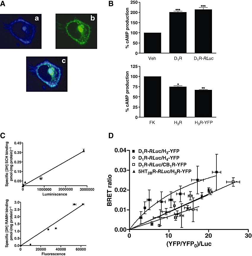
Experimental approach: D1-H3 receptor heteromerization was studied in mammalian transfected cells with Bioluminescence Resonance Energy Transfer and binding assays. Furthermore, signalling through mitogen-activated protein kinase (MAPK) and adenylyl cyclase pathways was studied in co-transfected cells and compared with cells transfected with either D1 or H3 receptors.
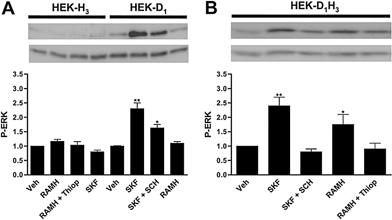
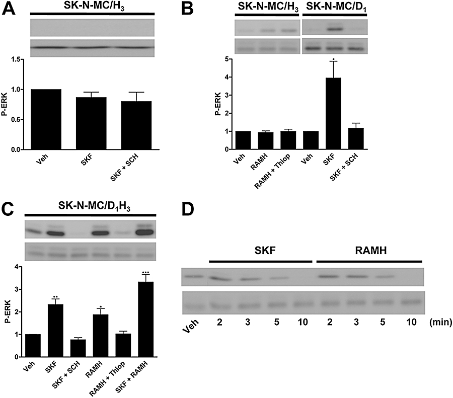
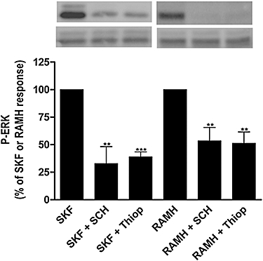
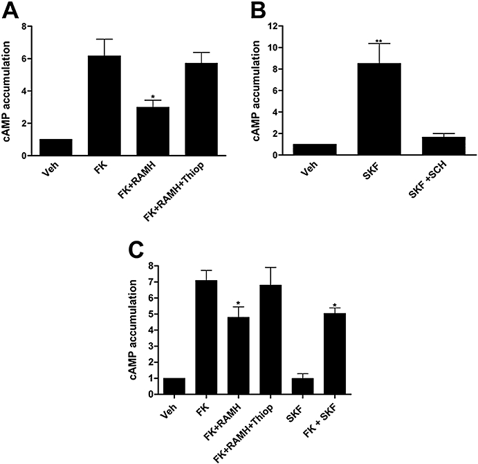
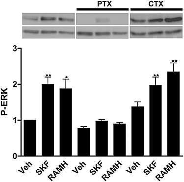
Key results: Bioluminescence Resonance Energy Transfer and binding assays confirmed that D1 and H3 receptors can heteromerize. Activation of histamine H3 receptors did not lead to signalling towards the MAPK pathway unless dopamine D1 receptors were co-expressed. Also, dopamine D1 receptors, usually coupled to G(s) proteins and leading to increases in cAMP, did not couple to G(s) but to G(i) in co-transfected cells. Furthermore, signalling via each receptor was blocked not only by a selective antagonist but also by an antagonist of the partner receptor.
Conclusions and implications: D1-H3 receptor heteromers constitute unique devices that can direct dopaminergic and histaminergic signalling towards the MAPK pathway in a G(s)-independent and G(i)-dependent manner. An antagonist of one of the receptor units in the D1-H3 receptor heteromer can induce conformational changes in the other receptor unit and block specific signals originating in the heteromer. This gives rise to unsuspected therapeutic potentials for G protein-coupled receptor antagonists.
Figures
Similar articles
-
Heteroreceptor Complexes Formed by Dopamine D1, Histamine H3, and N-Methyl-D-Aspartate Glutamate Receptors as Targets to Prevent Neuronal Death in Alzheimer's Disease.Mol Neurobiol. 2017 Aug;54(6):4537-4550. doi: 10.1007/s12035-016-9995-y. Epub 2016 Jul 1. Mol Neurobiol. 2017. PMID: 27370794
-
Dopamine D1-histamine H3 receptor heteromers provide a selective link to MAPK signaling in GABAergic neurons of the direct striatal pathway.J Biol Chem. 2011 Feb 18;286(7):5846-54. doi: 10.1074/jbc.M110.161489. Epub 2010 Dec 20. J Biol Chem. 2011. PMID: 21173143 Free PMC article.
-
Histamine H3 receptor activation prevents dopamine D1 receptor-mediated inhibition of dopamine release in the rat striatum: a microdialysis study.Neurosci Lett. 2013 Sep 27;552:5-9. doi: 10.1016/j.neulet.2013.07.026. Epub 2013 Jul 26. Neurosci Lett. 2013. PMID: 23896530
-
Modulation of neurotransmitter release via histamine H3 heteroreceptors.Fundam Clin Pharmacol. 1994;8(2):128-37. doi: 10.1111/j.1472-8206.1994.tb00789.x. Fundam Clin Pharmacol. 1994. PMID: 8020871 Review.
-
Progress in the development of histamine H3 receptor antagonists/inverse agonists: a patent review (2013-2017).Expert Opin Ther Pat. 2018 Mar;28(3):175-196. doi: 10.1080/13543776.2018.1424135. Epub 2018 Jan 15. Expert Opin Ther Pat. 2018. PMID: 29334795 Review.
Cited by
-
Human kidney-2 cells harbor functional dopamine D1 receptors that require Giα for Gq/11α signaling.Am J Physiol Renal Physiol. 2013 Aug 15;305(4):F560-7. doi: 10.1152/ajprenal.00644.2012. Epub 2013 May 22. Am J Physiol Renal Physiol. 2013. PMID: 23698121 Free PMC article.
-
Histamine-3 Receptor Availability and Glutamate Levels in the Brain: A PET-1H-MRS Study of Patients With Schizophrenia and Healthy Controls.Int J Neuropsychopharmacol. 2024 Mar 1;27(3):pyae011. doi: 10.1093/ijnp/pyae011. Int J Neuropsychopharmacol. 2024. PMID: 38373256 Free PMC article.
-
GPCR homomers and heteromers: a better choice as targets for drug development than GPCR monomers?Pharmacol Ther. 2009 Nov;124(2):248-57. doi: 10.1016/j.pharmthera.2009.07.005. Epub 2009 Aug 5. Pharmacol Ther. 2009. PMID: 19664655 Free PMC article. Review.
-
The histamine H3 receptor modulates dopamine D2 receptor-dependent signaling pathways and mouse behaviors.J Biol Chem. 2023 Apr;299(4):104583. doi: 10.1016/j.jbc.2023.104583. Epub 2023 Mar 4. J Biol Chem. 2023. PMID: 36871761 Free PMC article.
-
The Old and New Visions of Biased Agonism Through the Prism of Adenosine Receptor Signaling and Receptor/Receptor and Receptor/Protein Interactions.Front Pharmacol. 2021 Jan 29;11:628601. doi: 10.3389/fphar.2020.628601. eCollection 2020. Front Pharmacol. 2021. PMID: 33584311 Free PMC article.
References
-
- Agnati LF, Ferré S, Lluis C, Franco R, Fuxe K. Molecular mechanisms and therapeutical implications of intramembrane receptor/receptor interactions among heptahelical receptors with examples from the striatopallidal GABA neurons. Pharmacol Rev. 2003;55:509–550. - PubMed
-
- Agnati LF, Fuxe K, Ferré S. How receptor mosaics decode transmitter signals. Possible relevance of cooperativity. Trends Biochem Sci. 2005;30:188–193. - PubMed
-
- Anichtchik OV, Peitsaro N, Rinne JO, Kalimo H, Panula P. Distribution and modulation of histamine H(3) receptors in basal ganglia and frontal cortex of healthy controls and patients with Parkinson. s disease. Neurobiol Dis. 2001;8:707–716. - PubMed
Publication types
MeSH terms
Substances
Grants and funding
LinkOut - more resources
Full Text Sources
