

A spindle-independent cleavage furrow positioning pathway
- PMID: 20811457
- PMCID: PMC4028831
- DOI: 10.1038/nature09334
A spindle-independent cleavage furrow positioning pathway
Abstract
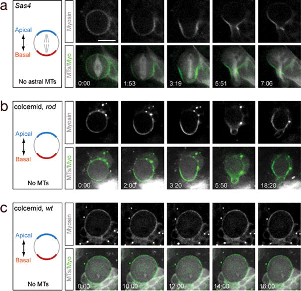
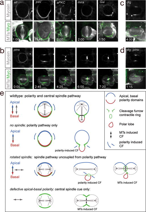
The mitotic spindle determines the cleavage furrow site during metazoan cell division, but whether other mechanisms exist remains unknown. Here we identify a spindle-independent mechanism for cleavage furrow positioning in Drosophila neuroblasts. We show that early and late furrow proteins (Pavarotti, Anillin, and Myosin) are localized to the neuroblast basal cortex at anaphase onset by a Pins cortical polarity pathway, and can induce a basally displaced furrow even in the complete absence of a mitotic spindle. Rotation or displacement of the spindle results in two furrows: an early polarity-induced basal furrow and a later spindle-induced furrow. This spindle-independent cleavage furrow mechanism may be relevant to other highly polarized mitotic cells, such as mammalian neural progenitors.
Figures


Similar articles
-
Asymmetrically dividing Drosophila neuroblasts utilize two spatially and temporally independent cytokinesis pathways.Nat Commun. 2015 Mar 20;6:6551. doi: 10.1038/ncomms7551. Nat Commun. 2015. PMID: 25791062 Free PMC article.
-
Inducing "cytokinesis" without mitosis in unfertilized Drosophila eggs.Cell Cycle. 2012 Aug 1;11(15):2856-63. doi: 10.4161/cc.21190. Epub 2012 Aug 1. Cell Cycle. 2012. PMID: 22801541
-
PAR-4 and anillin regulate myosin to coordinate spindle and furrow position during asymmetric division.J Cell Biol. 2015 Sep 28;210(7):1085-99. doi: 10.1083/jcb.201503006. J Cell Biol. 2015. PMID: 26416962 Free PMC article.
-
Mechanisms of Spindle Positioning: Lessons from Worms and Mammalian Cells.Biomolecules. 2019 Feb 25;9(2):80. doi: 10.3390/biom9020080. Biomolecules. 2019. PMID: 30823600 Free PMC article. Review.
-
Centralspindlin in Rappaport's cleavage signaling.Semin Cell Dev Biol. 2016 May;53:45-56. doi: 10.1016/j.semcdb.2016.03.006. Epub 2016 Mar 7. Semin Cell Dev Biol. 2016. PMID: 26964770 Review.
Cited by
-
The crosstalk between microtubules, actin and membranes shapes cell division.Open Biol. 2020 Mar;10(3):190314. doi: 10.1098/rsob.190314. Epub 2020 Mar 18. Open Biol. 2020. PMID: 32183618 Free PMC article. Review.
-
In Vivo Photocontrol of Microtubule Dynamics and Integrity, Migration and Mitosis, by the Potent GFP-Imaging-Compatible Photoswitchable Reagents SBTubA4P and SBTub2M.J Am Chem Soc. 2022 Mar 30;144(12):5614-5628. doi: 10.1021/jacs.2c01020. Epub 2022 Mar 15. J Am Chem Soc. 2022. PMID: 35290733 Free PMC article.
-
Asymmetric cortical extension shifts cleavage furrow position in Drosophila neuroblasts.Mol Biol Cell. 2011 Nov;22(22):4220-6. doi: 10.1091/mbc.E11-02-0173. Epub 2011 Sep 21. Mol Biol Cell. 2011. PMID: 21937716 Free PMC article.
-
Cortical PAR polarity proteins promote robust cytokinesis during asymmetric cell division.J Cell Biol. 2016 Jan 4;212(1):39-49. doi: 10.1083/jcb.201510063. J Cell Biol. 2016. PMID: 26728855 Free PMC article.
-
Mechanochemical Signaling Directs Cell-Shape Change.Biophys J. 2017 Jan 24;112(2):207-214. doi: 10.1016/j.bpj.2016.12.015. Biophys J. 2017. PMID: 28122209 Free PMC article. Review.
References
-
- Oliferenko S, Chew TG, Balasubramanian MK. Positioning cytokinesis. Genes Dev. 2009;23(6):660. - PubMed
-
- von Dassow G. Concurrent cues for cytokinetic furrow induction in animal cells. Trends Cell Biol. 2009;19(4):165. - PubMed
-
- Deng M, Suraneni P, Schultz RM, Li R. The Ran GTPase mediates chromatin signaling to control cortical polarity during polar body extrusion in mouse oocytes. Dev Cell. 2007;12(2):301. - PubMed
-
- Somers WG, Saint R. A RhoGEF and Rho family GTPase-activating protein complex links the contractile ring to cortical microtubules at the onset of cytokinesis. Dev Cell. 2003;4(1):29. - PubMed
Publication types
MeSH terms
Substances
Grants and funding
LinkOut - more resources
Full Text Sources
Molecular Biology Databases
Miscellaneous
