. 2019 May;180(1):289-304.
doi: 10.1104/pp.18.01225. Epub 2019 Feb 12.

The AAA-ATPase MIDASIN 1 Functions in Ribosome Biogenesis and Is Essential for Embryo and Root Development

Affiliations

The AAA-ATPase MIDASIN 1 Functions in Ribosome Biogenesis and Is Essential for Embryo and Root Development

Peng-Cheng Li et al. Plant Physiol. 2019 May.

Abstract

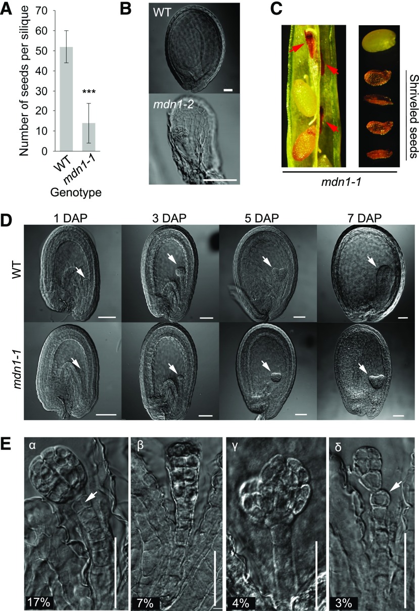
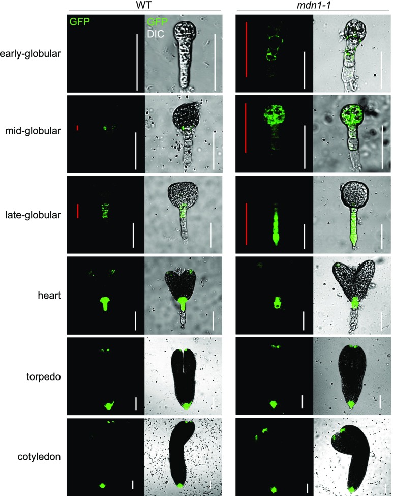
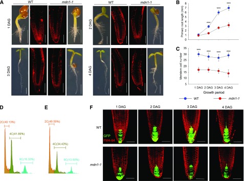
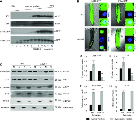
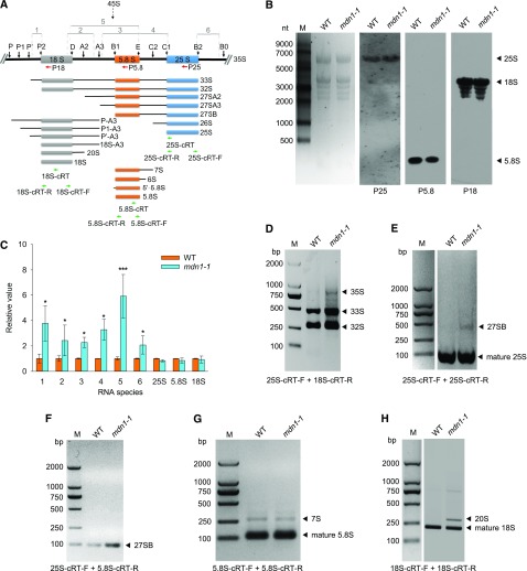
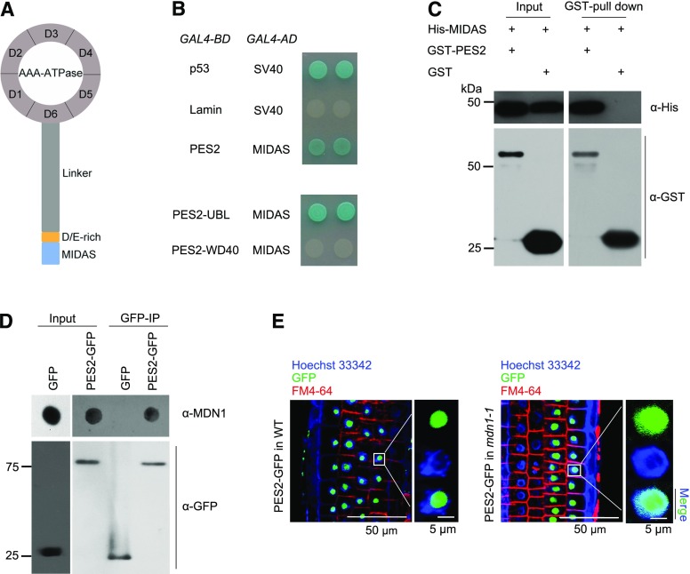
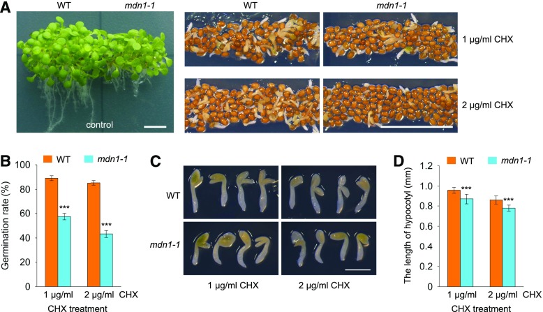
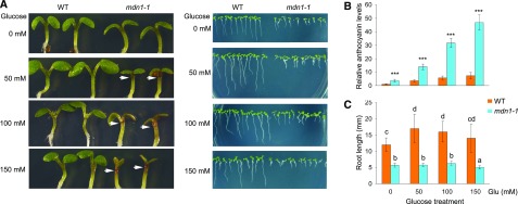
Ribosome biogenesis is an orchestrated process that relies on many assembly factors. The AAA-ATPase Midasin 1 (Mdn1) functions as a ribosome assembly factor in yeast (Saccharomyces cerevisiae), but the roles of MDN1 in Arabidopsis (Arabidopsis thaliana) are poorly understood. Here, we showed that the Arabidopsis null mutant of MDN1 is embryo-lethal. Using the weak mutant mdn1-1, which maintains viability, we found that MDN1 is critical for the regular pattern of auxin maxima in the globular embryo and functions in root meristem maintenance. By detecting the subcellular distribution of ribosome proteins, we noted that mdn1-1 impairs nuclear export of the pre-60S ribosomal particle. The processing of ribosomal precusor RNAs, including 35S, 27SB, and 20S, is also affected in this mutant. MDN1 physically interacts with PESCADILLO2 (PES2), an essential assembly factor of the 60S ribosome, and the observed mislocalization of PES2 in mdn1-1 further implied that MDN1 plays an indispensable role in 60S ribosome biogenesis. Therefore, the observed hypersensitivity of mdn1-1 to a eukaryotic translation inhibitor and high-sugar conditions might be associated with the defect in ribosome biogenesis. Overall, this work establishes a role of Arabidopsis MDN1 in ribosome biogenesis, which agrees with its roles in embryogenesis and root development.


Figures

Figure 1.
Figure 1.
Embryo developmental phenotypes of the mdn1 mutants. A, The number of plump seeds per silique of the wild type and mdn1-1. Error bars represent sd (n = 15). Student’s t test was applied (***P < 0.01). B, Embryos of the aborted seeds from the mdn1-2 siliques at 12 DAP observed with DIC optics. Scale bars = 50 μm. C, Shriveled seeds from the mdn1-1 siliques at 12 DAP. Red arrowheads indicate the abnormal seeds. D, Embryos of wild type (WT) and mdn1-1 at different developmental stages observed with DIC optics. White arrowheads indicate the embryos. Scale bars = 50 μm. E, Phenotypes of four types of malformed globular embryos of mdn1-1 at 4 DAP. The percentage of each type is indicated below. For the α-type, the white arrowhead indicates the abnormal cell division pattern. For the δ-type, the white arrowhead indicates the misshaped suspensor cell. Scale bars = 50 μm.
Figure 2.
Figure 2.
mdn1-1 embryos display altered auxin distribution. DR5rev::GFP expression in wild-type (WT) and mdn1-1 embryos at the indicated stages. Red lines indicate the distribution range of GFP signals in embryo. White lines are scale bars (= 50 μm). Images were acquired using the same optical setting.
Figure 3.
Figure 3.
Root development phenotypes of mdn1-1. A, Seedling and RAM phenotypes at the indicated stages grown under normal growth conditions. The images of seedlings were obtained with an optical microscope (scale bars = 1 mm). The images of FM4-64–stained RAM were obtained using a CLSM (scale bars = 20 μm). B, Kinematic analyses of primary root length of wild type and mdn1-1 from 1 to 4 DAG. C, Kinematic comparison of the root meristem cell number between wild type and mdn1-1 from 1 to 4 DAG. B and C, Error bars represent sd (n = 10), and Student’s t test was applied (***P < 0.01). D and E, Ploidy analysis of nuclei isolated from root cells of wild type and mdn1-1, respectively, using flow cytometry. Five independent experiments were performed, and representative results are presented. F, The expression pattern of DR5rev::GFP in root tips of wild type and mdn1-1 at the indicated stages. Scale bars = 20 μm. WT, wild type.
Figure 4.
Figure 4.
Nuclear export of pre-60S ribosomal particles is impaired in mdn1-1. A, Cofractionation analyses of L16B-GFP and S13A-GFP with 60S and 40S ribosomal subunits, respectively, using Suc gradient sedimentation and immunoblotting. The factions of 60S/80S and polysomes are indicated. L17 and S14 were employed as markers of 60S and 40S ribosomal subunits, respectively. B, Subcellular localization analyses of L16B-GFP and S13A-GFP in wild-type and mdn1-1 background. Nuclei were indicated by 4′,6-diamino-phenylindole staining. Images were acquired using the same optical setting. Scale bars = 100 µm (left images) and 5 µm (right images). C, Western-blot analyses of L16B-GFP, L17, S13A-GFP, and S14 in nuclear (N) and cytoplasmic (C) fractions extracted from 5-d-old wild-type (WT) and mdn1-1 seedlings. Each lane was loaded with 10-μg protein. Six biological replicates were performed, and representative results are presented. D–G, The protein levels of L16B-GFP (D), L17 (E), S13A-GFP (F), and S14 (G) in the cytoplasmic fraction relative to that in the nuclear fractions of wild type (WT) and mdn1-1. The protein levels were according to gray values of the western-blot bands. Error bars represent sd (n = 6), and Student’s t test was applied (***P < 0.01).
Figure 5.
Figure 5.
PrerRNA processing in mdn1-1. A, Diagram of the rRNAs and pathways of prerRNA processing in Arabidopsis. The major processing intermediates are illustrated. The black vertical arrows indicate the processing sites. Regions 1–6 indicate the fragments that are employed for RT-qPCR amplification. The regions used for oligonucleotide probes are indicated with red horizontal arrows. The regions of the primers used for reverse transcription and cRT-PCR amplification are indicated with green horizontal arrows. B, Northern-blot analyses of the mature rRNA levels in 5-d-old seedlings of wild type and mdn1-1 with specific probes indicated in (A). M, RNA size marker. Left, the ethidium-bromide–stained gel image, which is shown as a loading control. C, Levels of the intermediates indicated in (A) and three mature rRNAs in mdn1-1 relative to that in wild type. Arabidopsis Actin7 was employed as the internal control to normalize the values. Error bars represent sd (n = 4). Student’s t test was applied (***P < 0.01, and *P < 0.05). D, Increased accumulation of the 35S, 33S, and 32S prerRNAs in mdn1-1 determined by PCR with 25S-cRT-F and 18S-cRT-R primers. E and F, Increase of the 27SB intermediate in mdn1-1 validated by PCR with 25S-cRT-F and 25S-cRT-R primers (E) and 25S-cRT-F and 5.8S-cRT-R primers (F). G, Unchanged levels of the 7S prerRNA and 5.8S rRNA in mdn1-1 checked by PCR with 5.8S-cRT-F and 5.8S-cRT-R primers. H, Increased accumulation of the 20S intermediate in mdn1-1 determined by PCR with 18S-cRT-F and 18S-cRT-R primers. D–H, Bands indicated by arrowheads were cloned and sequenced. M, DNA markers; WT, wild type; bp, basepair.
Figure 6.
Figure 6.
The interaction between MDN1 and PES2 in vitro and in vivo. A, Illustration of the MDN1 protein domains. Ring-shaped AAA-ATPase consists of six tandem AAA protomers (D1–D6), a long flexible linker, a D/E-rich domain, and the MIDAS domain at the tip. B, Yeast-2-hybrid assays on protein interaction between MIDAS and PES2, the UBL-domain of PES2 (PES2-UBL), or the WD40-domain of PES2 (PES2-WD40). Plasmids expressing the indicated GAL4-BD and GAL4-AD were transformed into the yeast strain Y2HGold. The interaction of p53 and the SV40 large T-antigen was used as the positive control, while that of lamin and SV40 was used as the negative control. Solutions of transformed yeast were spotted onto the SD/-Ade/-His/-Leu/-Trp/X-α-Gal/AbA medium. C, GST-tagged PES2 interacts with the His-6-tagged MIDAS in a GST pull-down assay. D, IP analysis of PES2-GFP with the MDN1 protein. Protein extracts from the homozygous transgenic seedlings of 35S::PES2-GFP and 35S::GFP were subjected to IP with the monoclonal anti-GFP antibody. The MDN1 protein was detected by dot-immunoblot with the anti-MDN1 antibody. E, Subcellular localization of PES2-GFP in RAM cells of 4-d-old seedling of the wild-type (WT) or mdn1-1 background. The nucleoplasm was indicated with the Hoechst 33342 staining, and the cellular outline was indicated with the FM4-64 dye. Scale bars = 50 µm (left images) and 5 µm (right images).
Figure 7.
Figure 7.
Phenotypes of mdn1-1 grown under CHX-treatment conditions. A, Phenotypes of wild type and mdn1-1 grown on solid 1/2 MS medium containing 0 (control), 1 μg/mL, or 2 μg/mL CHX for 2 weeks. Scale bars = 50 mm. B, Germination rate analyses of wild type and mdn1-1 grown under the indicated CHX-treatment conditions. Data are mean values of three biological replicates ± sd. Student’s t test was applied (***P < 0.01). C, Seedling phenotypes of wild type and mdn1-1 grown under CHX-treatment conditions for 2 weeks. Scale bar = 1 mm. D, Comparison analyses of the length of hypocotyl with radicle between wild type and mdn1-1 grown under CHX-treatment conditions for 2 weeks. Error bars = sd (n = 10). Student’s t test was applied (***P < 0.01). WT, wild type.
Figure 8.
Figure 8.
Phenotypes of mdn1-1 grown under Glc-treatment conditions. A, Phenotypes of wild type and mdn1-1 grown on solid MS medium containing the indicated concentration Glc for 5 d. The white arrowheads indicate the accumulated anthocyanins. B, Relative anthocyanin levels in the seedlings shown in (A). The anthocyanin content of wild type grown on Glc-free medium was set to 1. Error bars = SD (n = 3). Student’s t test was applied (***P < 0.01). C, Comparison analyses of root length of seedlings shown in (A). Data are mean values of 10 replicates ± sd. Statistically significant differences are indicated by different lowercase letters (P < 0.01, Duncan's multiple range test). WT, wild type.

Similar articles

Cited by

References

    1. Abbasi N, Kim HB, Park NI, Kim HS, Kim YK, Park YI, Choi SB (2010) APUM23, a nucleolar Puf domain protein, is involved in pre-ribosomal RNA processing and normal growth patterning in Arabidopsis. Plant J 64: 960–976 - PubMed
    1. Ahn CS, Cho HK, Lee DH, Sim HJ, Kim SG, Pai HS (2016) Functional characterization of the ribosome biogenesis factors PES, BOP1, and WDR12 (PeBoW), and mechanisms of defective cell growth and proliferation caused by PeBoW deficiency in Arabidopsis. J Exp Bot 67: 5217–5232 - PMC - PubMed
    1. Barrio-Garcia C, Thoms M, Flemming D, Kater L, Berninghausen O, Bassler J, Beckmann R, Hurt E (2016) Architecture of the Rix1-Rea1 checkpoint machinery during pre-60S-ribosome remodeling. Nat Struct Mol Biol 23: 37–44 - PubMed
    1. Bassler J, Kallas M, Pertschy B, Ulbrich C, Thoms M, Hurt E (2010) The AAA-ATPase Rea1 drives removal of biogenesis factors during multiple stages of 60S ribosome assembly. Mol Cell 38: 712–721 - PMC - PubMed
    1. Bassler J, Paternoga H, Holdermann I, Thoms M, Granneman S, Barrio-Garcia C, Nyarko A, Lee W, Stier G, Clark SA, et al. (2014) A network of assembly factors is involved in remodeling rRNA elements during preribosome maturation. J Cell Biol 207: 481–498 - PMC - PubMed

Publication types

MeSH terms

LinkOut - more resources